
Radiographs (x-ray)
Diagnostics are a crucial tool in veterinary care that we employ at Hanover Animal Hospital to assist diagnose our animals’ conditions.

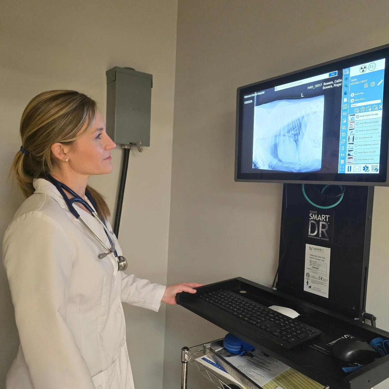
Diagnostic Imaging
With the advancements in digital X-ray technology, we can better determine what is wrong. This has made it possible for us to find things like hairline fractures and previously invisible orthopedic disorders. Specialists who consult with us on challenging cases receive access to these digital photographs. The use of radiology (x-rays) to examine a pet’s bones, digestive system (stomach, intestines, colon), respiratory system (lungs), heart, and genitourinary system often yields important information (bladder, prostate). It can be used independently or in conjunction with other diagnostic techniques to pinpoint the precise source of a problem, rule out potential issues, or provide a list of potential causes for a pet’s condition.